


|
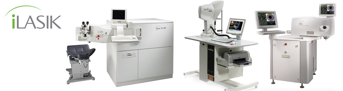
Microkeratome Blades and Corneal Flap |
|
Chi-Wang Yau, MD; Huey-Chuan Cheng, MD, MSc |
|
丘子宏眼科診所 丘 子 宏
|
|
馬偕紀念醫院眼科 鄭 惠 川
丘子宏院長雖然自行開業十多年,但在繁忙的門診及手術中,仍不忘醫學研究的工作。本篇論文被國際眼科醫學雜誌「Ophthalmic Surgery Laser & Imaging」接受刊登,是非常不容易的。
研究目的:在雷射手術中,所謂 “ LASIK”,最重要的第一個步驟是在角膜以微切刀 ( Microkeratome) 切取角膜瓣,角膜瓣切取是決定接下來能否用雷射光矯正近視的關鍵步驟。因為如果角膜瓣切得太厚,接下來就沒有足夠的角膜厚度可以雷射了。如果勉強去雷射,角膜的剩餘厚度不足,將有產生「圓錐角膜」的危險。相反的,如果切得太薄,又有角膜瓣穿孔、縐褶的併發症。
丘院長經多年來實行術中測量角膜厚度的技術「Intra-operative Pachymetry」,發現微切刀切取的角膜瓣厚度,誤差相當大,除了眾所週知與所用的微切刀種類有關外,發現所用的刀片也很有非常重要的影響。因此,要切出理想厚度的角膜瓣,必須選擇正確的微切刀和刀片。
|
[Ophthalmic Surg Lasers Imaging 2008;39:471-475.]
INTRODUCTION
______________________________________________________________
From the Department of Ophthalmology (C-WY, H-CC), Mackay Memorial ______________________________________________________________
PATIENTS AND METHODS
The LASIK procedures were performed under topical anesthesia with 0.5% proparacaine eye drops given 3 minutes before and again just before insertion of the eye speculum. Under an operating microscope, excessive
For the first 2 years that we used the Moria microkeratomemicrokeratome, we also used Moria blades (Moria, France). We then switched to CLB blades (Med-Logics Inc., Laguna Hills, CA). We retrospectively analyzed the corneal flap thickness in the last 100 consecutive patients using the Moria blades and the first 100 consecutive patients using the CLB blades. The study group thus comprised a total of 200 patients and 400 eyes. The Student’s t test was used to compare the corneal thickness in the Moria and CLB groups and also in the first and second eyes of all patients. Data are reported as mean ± standard deviation (SD).
RESULTS
DISCUSSION
Several investigators have studied the flap thickness produced by the Moria M2 microkeratome (Table 3).5,6,8,12,13 Solomon et al.8 reported the mean flap thicknesses using the 130 head were 150 and 148 μm in the first and second eyes. Miranda et al.5 used a 110 head in 257 eyes, obtaining a mean flap thickness of 134 μm. Muallem et al.6 found that the 110 head with fast translation velocity created a 136- and 132-μm flap in the first and second eye, respectively. The mean flap thicknesses in those studies were considered close to the desire thicknesses. However, Muallem et al. also found that the head translation velocity significantly affected the flap thickness. With a 110 head with a slow translation velocity, they obtained much thicker flaps of 151 and 148 μm in the first and second eyes, comparable to those produced by the 130 head.
The current investigation demonstrated that the choice of blade alone may also affect flap thickness. All other conditions for the LASIK procedure were the same in both groups of patients. Using the same Moria M2 110 head, we found that Moria blades created a significantly thicker flap than did CLB blades. The flap thickness in our series using Moria blades is similar to the results of other studies,5,6 but we were unable to find reports comparing it with the CLB blades. Therefore, one must use the same brand of blades to obtain consistent flap thickness. Alternatively, if changing blade brands for any reason, one should be aware that the results obtained may be different than expected. Our study was not designed to examine why different brands of blades produce different flap thicknesses. Factors such as the design, sharpness, and material of the blades might contribute to the variable performance. This issue needs to be examined from an engineering perspective.
We found that the Moria blade yields a wider range of flap thickness than the CLB blade (SD = 21.4 and 21.6 in the first and second eyes for the Moria blade vs 12.1 and 15.5 for the CLB blade). Muallem et al.6 found similar results with the Moria M2 microkeratome (SD = 23.5 to 25.5), but they did not specify what blade(s) they used. This is comparable to results reported with other microkeratomes. The smallest reported SD (13.3) was for the Nidek MK-2000 microkeratome.14 Interestingly, we found a smaller SD with the CLB blades, especially in the first eyes (Table 4). As a goal for future microkeratome designs, Choudhri et al.15 proposed that the mean SD be reduced to below 10 μm. In our hands, the CLB blade in a Moria M2 microkeratome did not achieve that goal, but it came closer to it than the Moria blade.
If the same blade is used for both eyes, there is a tendency to cut a thinner flap in the second eye.5,8,16-18 Muallem et al.6 found this was true with the Moria M2 130 head but not so much with the 110 head. The reason for a thinner flap in the second eye is not clear, but most authors suggest it is because the blade is dulled when cutting the first eye.6,19 We found a significantly thinner second flap cut with the CLB blades, but there was no significant difference with the Moria blades. This again demonstrates different performance by different brands of blades during LASIK surgery.
The thinner flap cut by the CLB blades may be helpful in treating patients with a thin cornea and high degree myopia. With the thin flap, one can leave thicker stromal tissue to correct a high refractive error. The difference in flap thickness might also be exploited in particular cases. For example, one could deliberately choose the more myopic eye as the second eye, expecting a thinner flap in that eye. There are therefore potential benefits to be derived from the characteristic results of the CLB blade. At the same time, this must be balanced against the risk of an excessively thin or even a buttonhole flap for the second eye. It might be wise to use intraoperative pachymetry to verify the flap thickness. If the flap in the first eye is relatively thin, a new blade for the second eye should be used to avoid such complications. The current study showed that the corneal flap thickness produced by a Moria M2 microkeratome 110 head is variable depending on the blade used. CLB blades produced significantly thinner flaps than Moria blades. This study therefore adds to the list of factors potentially affecting flap thickness. Surgeons performing LASIK should be aware of this factor and be cautious when using a different type of blade than they have had experience with. Under such circumstances, we recommend using intraoperative pachymetry to evaluate corneal flap thickness until one is familiar with the results yielded by the new type of microkeratome blade.
REFERENCES
2. Pallikaris IG, Kymionis GD, Astyrakakis NI. Corneal ectasia induced by laser in situ keratomileusis. J Cataract Refract Surg. 2001;27:1796-1802.
3. Durairaj VD, Balentine J, Kouyoumdjian G, et al. The predictability of corneal flap thickness and tissue laser ablation in laser in situ keratomileusis. Ophthalmology. 2000;107:2140-2143.
4. Jacobs BJ, Deutsch TA, Rubenstein JB. Reproducibility of corneal flap thickness in LASIK. Ophthalmic Surg Lasers. 1999;30:350-353.
5. Miranda D, Smith SD, Krueger RR. Comparison of flap thickness reproducibility using microkeratomes with a second motor for advancement. Ophthalmology. 2003;110:1931-1934.
6. Muallem MS, Yoo SY, Romano AC, Schiffman JC, Culbertson WW. Corneal flap thickness in laser in situ keratomileusis using the Moria M2 microkeratome. J Cataract Refract Surg. 2004;30:1902-1908.
7. Naripthaphan P, Vongthongsri A. Evaluation of the reliability of the Nidek MK-2000 microkeratome for laser in situ keratomileusis. J Refract Surg. 2001;17:S255-S258.
8. Solomon KD, Donnenfeld E, Sandoval HP. Flap thickness accuracy: comparison of 6 microkeratome models. J Cataract Refract Surg. 2004;30:964-977.
9. Yi WM, Joo CK. Corneal flap thickness in laser in situ keratomileusis using an SCMD manual microkeratome. J Cataract Refract Surg. 1999;25:1087-1092.
10. Yildirim R, Aras C, Ozdamar A, Bahcecioglu H, Ozkan S. Reproducibility of corneal flap thickness in laser in situ keratomileusis using the Hansatome microkeratome. J Cataract Refract Surg. 2000;26:1729-1732.
11. Buratto L, Brint S, Salib G. LASIK with a nasal hinge. In: Buratto L, Brint S, eds. Custom LASIK: Surgical Techniques and Complications. Thorofare, NJ: SLACK Incorporated; 2003:93-111.
12. Guell JL, Velasco F, Roberts C, Sisquella MT, Mahmoud A. Corneal flap thickness and topography changes induced by flap creation during laser in situ keratomileusis. J Cataract Refract Surg. 2005;31:115-119.
13. Javaloy JE, Quinto A, De Rojas V, Alio JL. Quality assessment model of 3 different microkeratomes through confocal microscopy. J Cataract Refract Surg. 2004;30:1300-1309.
14. Sarkisian KA, Petrov AA. Experience with the Nidek MK-2000 microkeratome in 1,220 cases. J Refract Surg. 2001;17:S252-S254.
15. Choudhri SA, Feigenbaum SK, Pepose JS. Factors predictive of LASIK flap thickness with the Hansatome zero compression microkeratome. J Refract Surg. 2005;21:253-259.
16. Gailitis RP, Lagzdins M. Factors that affect corneal flap thickness with the Hansatome microkeratome. J Refract Surg. 2002;18:439-443.
17. Jackson DW, Wang L, Koch DD. Accuracy and precision of the amadeus microkeratome in producing LASIK flaps. Cornea. 2003;22:504-507.
18. Shemesh G, Dotan G, Lipshitz I. Predictability of corneal flap thickness in laser in situ keratomileusis using three different microkeratomes. J Refract Surg. 2002;18: S347-S351. 19. Viestenz A, Langenbucher A, Hofmann-Rummelt C, Modis L, Viestenz A, Seitz B. Evaluation of corneal flap dimensions and cut quality using the SKBM automated microkeratome. J Cataract Refract Surg. 2003;29:825-831. |